Roger McLendon on Solving the Jigsaw Puzzle of Glioblastoma
Special Topic of Glioblastoma Interview, October 2011

|
Our Special Topics analysis of Glioblastoma research over the past decade shows that the work of Dr. Roger McLendon ranks at #5 by total cites and #12 by total papers, based on 73 papers cited a total of 5,311 times during the analysis period. Four of these papers rank among the 20 most-cited over the past decade or over the past two years. In Essential Science IndicatorsSM from Clarivate, McLendon ranks in the top 1% among scientists in the field of Clinical Medicine. McLendon is Chief of Neuropathology and Surgical Pathology, Director of Anatomic Pathology Services, and Director of the Preston Robert Tisch Brain Tumor Center Tissue Bank at Duke University Medical Center in Durham, North Carolina. |
Below, he talks with ScienceWatch.com about his highly cited work as it relates to glioblastoma.
![]() Please tell us about your educational
background and research experiences.
Please tell us about your educational
background and research experiences.
I first became interested in the neurosciences during a project that I did at Emory Medical School when I was an undergraduate at Emory in Atlanta. The project involved tracing axonal pathways using silver stains. From there, I went to Medical College of Georgia where I was heavily influenced by Dr. Holde Puchtler and Dr. Dale Sickles, histochemists who were very successful at persuading a section of tissue to give up more information about its biology than what could be seen by the standard hematoxylin and eosin stain.
From there I went to Duke where I worked with Drs. Peter Burger, Darell Bigner, and Stephen Vogel, all outstanding neuropathologists and scientists. It was under them that I learned how to bring the findings from the basic science lab to the anatomic pathology lab.
![]() What first drew you to work with brain tumor
pathology in particular?
What first drew you to work with brain tumor
pathology in particular?
During my senior year in medical school, my main interest in neuropathology was in muscle diseases, but Dr. Sam Robinson, a neurosurgeon, advised me that if I was going to make a living in neuropathology, I would have to know something about brain tumors. He encouraged me to look into the epidemiological data available from the Georgia Tumor Registry to see what I could learn. This became the source data of my first paper, "The Glioblastoma Multiforme in Georgia, 1977-1981" (McLendon RE, et al., Cancer 56[4]: 894-7, 1985), and I never looked back.
At Duke, Dr. Peter Burger noted my interest in histochemistry and introduced me to Dr. Darell Bigner, with whom I described the diagnostic applications of the first monoclonal antibodies to glial fibrillary acidic protein in astrocytomas. Since that first project in 1984, I have collaborated continuously with Darell, Dr. Allan Friedman, and Dr. Henry Friedman at the Duke Brain Tumor Center as neuropathologist and, in 2000, I took over directorship of the Brain Tumor Center's biorepository. This collaborative nature has been something that both Darell and Henry have promoted and insisted on since I have been at Duke and has resulted in some very exciting clinical and basic science discoveries over the years.
![]() Your most-cited original article is the 2006
Nature paper you coauthored, "Glioma
stem cells promote
radioresistance by preferential activation of the DNA damage response,"
(Bao SD, et al., 444[7120]: 756-60, 7 December 2006). Would you
tell us a bit about this paper—your expectations going in, your
findings, where this work has gone since this publication?
Your most-cited original article is the 2006
Nature paper you coauthored, "Glioma
stem cells promote
radioresistance by preferential activation of the DNA damage response,"
(Bao SD, et al., 444[7120]: 756-60, 7 December 2006). Would you
tell us a bit about this paper—your expectations going in, your
findings, where this work has gone since this publication?
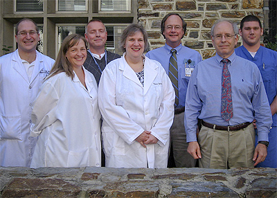
Pictured left to right: Dr. Eric Lipp, Lisa Ehinger,
Michael Leonard, Diane Satterfield, Dr. McLendon, Robert Annechiarico, and
Benjamin Wiener.
The great aspect of working at Duke has been the collaborative nature of the research. As a neuropathologist, I find myself "looking over the shoulders" of a lot of really bright people. They come up with a really great idea or finding and want to know how it applies to what we see in the microscope. Around 2000, Dr. Jeremy Rich joined the neuro-oncology faculty of the Brain Tumor Center and brought with him a great curiosity about the complex biology of the gliomas.
His desire was to dissect out the individual interactions among the several cell types in the tumor. We had known that glioblastomas exhibited a profound heterogeneity with respect to antigenic expression since Darell Bigner had written the key descriptive paper back in the '70s. The problem all of us are faced with is where does the heterogeneity come from and how is the diversity managed in the context of a growing tumor?
Jeremy started out by trying to genetically engineer astrocytes in cell culture to become immortal, producing the paper, "A genetically tractable model of human glioma formation," (Rich JN, et al., Cancer Research 61[9]: 3556-60, 1 May 2001). This also drove his curiosity about glioma cell invasion which he was pursuing through SPARC and bone morphogenetic protein manipulation. Jeremy and I began to discuss his findings and talk about brain tumor histology and the resemblance of his models to reality as seen in human gliomas.
About this time, Peter Dirks had described using the cell surface antigen, CD133, to localize glioma cells with stem cell properties. Jeremy began to work with the idea that these primitive stem cell-like glioma cells may provide the nidus for neoplastic progression, an idea that was similar to the ideas pushed by Kernohan in the '40s. However, it was not just that this subset of tumor cells had a growth advantage over the other cells, it became apparent through xenografting experiments that these stem cells were inducing their non-stem cell neoplastic, and even non-neoplastic neighbors, to proliferate and to migrate. Thus in a few experiments, he laid out a whole new avenue of research that has kept the whole collaborative team busy.