Steven Finkbeiner on Understanding Causes of Neurodegenerative Disease
Emerging Research Front Commentary, December 2010

|
Article: Inclusion body formation reduces levels of mutant huntingtin and the risk of neuronal death
Authors: Arrasate, M;Mitra, S;Schweitzer, ES;Segal,
MR;Finkbeiner, S |
Steven Finkbeiner talks with ScienceWatch.com and answers a few questions about this month's Emerging Research Front paper in the field of Neuroscience & Behavior.
![]() Why do you think your paper is highly
cited?
Why do you think your paper is highly
cited?
It provided a surprising and unequivocal answer to a longstanding debate about the role of abnormal protein deposits in neurodegeneration.
![]() Does it describe a new discovery, methodology, or
synthesis of knowledge?
Does it describe a new discovery, methodology, or
synthesis of knowledge?
It describes the invention of a new methodology, robotic microscopy, which was required to discover the role of protein deposition in neurodegeneration.
![]() Would you summarize the significance of your paper
in layman's terms?
Would you summarize the significance of your paper
in layman's terms?
Abnormal intracellular and extracellular protein deposits in the brain are pathological hallmarks of major neurodegenerative diseases, including Alzheimer's disease, Parkinson's disease, amyotrophic lateral sclerosis, and Huntington's disease. Since the discovery of these deposits, their role in disease has been debated. The prevailing view was that they are the principal cause of neurodegenerative disease, but some reports suggested that they were incidental or even represented a coping response.
To resolve the issue, we invented a new form of imaging called robotic microscopy and longitudinal analysis. We built an automated microscope and created software that enables it to find and return to the same individual neuron as often and for as long as the user wants, over weeks to months for example. With this microscope, the lives of thousands of individual neurons can be reconstructed through a series of images.
In turn, these longitudinal datasets can be analyzed with powerful statistical tools, known as survival analysis or Cox proportional hazards analysis. Commonly used in clinical or engineering studies, these tools make it possible to measure the extent to which an identified factor predicts an outcome of interest.
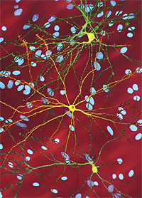
This figure illustrates a striatal neuron (yellow) that
harbors the mutant huntingtin protein, which has formed an inclusion body
(red). In the paper being featured by ScienceWatch.com, we show
that inclusion body formation is a coping response rather than a pathogenic
one, as it has been assumed.
Download additional
figures and descriptions of Martin Ostoja-Starzewski's work.
We applied this new technology to a primary neuron model of Huntington's disease. We observed the mutant protein as it was first expressed, determined whether or not a protein deposit called an inclusion body (IB) formed, and whether and when the neuron died. IB formation was quickly followed by a rapid reduction in levels of mutant protein elsewhere in the neuron and was not required for the mutant protein to cause neurodegeneration.
Surprisingly, neurons that formed IBs lived longer than those that did not. In fact, IB formation rapidly reduced the risk of cell death to near baseline levels. Instead, levels of other forms of the mutant protein elsewhere in the neuron predicted whether and when a neuron died.
The significance of the paper is twofold. First, it described a new method, robotic microscopy and longitudinal analysis, to determine whether disease-associated changes are pathogenic, incidental, or coping responses. Second, it showed that IB formation is likely a coping mechanism that reduces levels of the more toxic diffuse forms of the protein instead of a pathogenic process, as it was presumed to be.
![]() How did you become involved in this research, and
how would you describe the particular challenges, setbacks, and
successes that you've encountered along the way?
How did you become involved in this research, and
how would you describe the particular challenges, setbacks, and
successes that you've encountered along the way?
I am a neuroscientist and a neurologist, so I knew the importance of understanding causes of neurodegenerative disease. I took a particular interest in Huntington's disease on the advice of a mentor, Dr. Joseph Martin. I became aware of the limitations of conventional approaches for studying neurodegenerative disease during my postdoctoral fellowship in cellular and molecular biology with Dr. Michael Greenberg at Harvard.
The realization that conventional approaches would never be able to unequivocally resolve a whole class of fundamental questions in disease-related neuroscience was a powerful motivation for me. The more I contemplated the limitations of these approaches, the more I realized that unless I did something, I could spend my career generating results that might be wrong.
Finding a new way to deal with this limitation was my goal as I started my independent laboratory at the Gladstone Institutes and the University of California San Francisco. Fortunately, I had received training in computer programming, mathematics, and imaging from my graduate school mentors, Drs. Charles Stevens and Stephen Smith. Those skills enabled me to invent robotic microscopy and longitudinal analysis.
The main challenge here was not much different than most high-risk high-reward projects in academia. Funding agencies didn't believe that the invention was possible until it was invented, so I had to bet my start-up funds and a gift from a philanthropist that it would work.
Time spent developing the method meant time not publishing papers, and as a junior faculty member, I was under tremendous pressure from my Institution to publish and get grant support. The potential of the invention was hard for colleagues to grasp until it was demonstrated in this paper, so I didn't receive much encouragement. The institution made it clear that the risk I was taking was real. With a family to support, it was a challenge.
Fortunately, a talented postdoctoral fellow, Montserrat Arrasate, had the courage to work on this risky project with me. Nature sent the paper out for review, but then rejected it because of a very negative third reviewer. We really believed in the work, so we wrote a 14-page response to the reviews. Thankfully, the editor reconsidered the paper on appeal.
In the end, after another round of reviews and revisions, things turned out OK. The paper was published in an issue coinciding with the annual Society for Neuroscience meeting and was featured on the cover and in a News & Views piece by Dr. Harry Orr.
![]() Where do you see your research leading in the
future?
Where do you see your research leading in the
future?
The discovery that IB formation might be a coping response in a neuron model of Huntington's disease led to a re-evaluation of protein deposits in other neurodegenerative diseases. We subsequently developed primary neuron models of Parkinson's disease and amyotrophic lateral sclerosis/frontotemporal dementia and found that IBs are not required for neurodegeneration in these models either.
Rather than being a common pathogenic thread, these results suggest that IB formation is a common neuronal mechanism to cope with proteins prone to misfolding. The discovery also led to a renewed appreciation of the significant coping mechanisms that neurons mount in these diseases, which might explain why people don't usually become symptomatic until later in life.
"Abnormal intracellular and extracellular protein deposits in the brain are pathological hallmarks of major neurodegenerative diseases, including Alzheimer's disease, Parkinson's disease, amyotrophic lateral sclerosis, and Huntington's disease."
Therapeutic approaches traditionally focus on inhibiting pathogenic mechanisms, but this discovery suggests that the elucidation of coping mechanisms could lead to the identification of new targets whose pharmacological induction might be therapeutic.
The method of robotic microscopy and longitudinal analysis has proven to be extremely powerful, about a 100- to 1,000-fold more sensitive than the snapshot-based approaches that we used to use. The repeated longitudinal nature of the measurements substantially reduces the variability associated with cell-based assays, and it has enabled us to build later-generation systems adapted for high-throughput screening on small populations of heterogeneous primary cells, such as differentiated induced pluripotent stem cells. So, in addition to hypothesis-driven research, we are now able to do unbiased RNAi and small-molecule screens, for example, in primary cells.
Lastly, the mathematics of longitudinal analysis makes it possible to construct predictive, dynamical, multiparametric probabilistic models of biological or pathobiological processes. The quantitative nature of this approach is such that it can tell whether a factor tends to promote or retard an outcome of interest and also provides some measure of its overall importance. In effect, it allows us to develop dynamical "systems" models of health and disease.
The accuracy with which we can predict fate with the models we construct is a measure of how well we understand the underlying processes. The development of accurate quantitative predictive models of neurodegenerative disease could be fundamental for delineating successful therapeutic strategies.
![]() Do you foresee any social or political
implications for your research?
Do you foresee any social or political
implications for your research?
Neurodegenerative diseases are the sixth leading killer in the US, and the incidence is expected to increase with the rapid aging of populations in the US, Europe, Japan, and China. Despite the societal burden of these diseases, not a single disease-modifying therapy exists. Sadly, industry views neurodegenerative disease as a high-risk investment. Consequently, there are few promising treatments in the pipeline.
Hopefully, the discovery and approaches in this paper will help companies avoid mistaking coping responses for pathogenic ones and help redirect resources to targets that are likely to make a big difference in neurodegenerative disease. The technology might also enable primary screening in primary human cells, which may lead to the identification of therapeutics that have a better chance of being clinically safe and effective.
If the technology can significantly reduce the risk of identifying targets
and developing therapeutics for neurodegenerative disease, it could lead to
a virtuous cycle of increased success in clinical trials and increased
investment. The development of safe and effective therapies for
neurodegenerative diseases would have major societal
implications.![]()
![]() Steven Finkbeiner, M.D., Ph.D.
Steven Finkbeiner, M.D., Ph.D.
Professor, Departments of Neurology and Physiology
University of California, San
Francisco
Senior Investigator and Associate Director, Gladstone Institute of
Neurological Disease
Director, Taube-Koret Center for Huntington's Disease
Research
San Francisco, CA, USA
KEYWORDS: GREEN FLUORESCENT PROTEIN; INTRANUCLEAR INCLUSIONS; NEURODEGENERATIVE DISEASES; POLYGLUTAMINE EXPANSIONS; NEUROLOGICAL PHENOTYPE; TRANSGENIC MICE; IN-VITRO; AGGREGATION; NUCLEAR; REPEAT.