Jan Scholz Talks About Training the Brain
New Hot Paper Commentary, March 2011


|
Article: Training induces changes in white-matter architecture
Authors: Scholz, J;Klein, MC;Behrens,
TEJ;Johansen-Berg, H |
Jan Scholz talks with ScienceWatch.com and answers a few questions about this month's New Hot Paper in the field of Neuroscience & Behavior.
![]() Why do you think your paper is highly cited? Does
it describe a new discovery, methodology, or synthesis of knowledge?
Would you summarize the significance of your paper in layman's terms?
How did you become involved in this research, and how would you
describe the particular challenges, setbacks, and successes that
you've encountered along the way?
Why do you think your paper is highly cited? Does
it describe a new discovery, methodology, or synthesis of knowledge?
Would you summarize the significance of your paper in layman's terms?
How did you become involved in this research, and how would you
describe the particular challenges, setbacks, and successes that
you've encountered along the way?
In our work we presented evidence that the adult brain changes in response to a relatively short period of juggling practice. With non-invasive magnetic resonance imaging we imaged the brain of a group of healthy adult volunteers before they started practicing and following the six-week training period. We found evidence for structural changes in cortical areas and underlying connections. Our work was the first to suggest that such changes occur in the adult brain’s connections after motor training.
Our work followed up on a popular research subject previously examined by Eleanor A. Maguire (2000), Bogdan Draganski (2004), and Sara L. Bengtsson (2005): Do experience and practice influence brain structure? All of our studies have been highly cited and have received a lot of press coverage and interest from the general public. The previous studies, however, have only examined one brain region at a time. They either looked at the brain’s grey matter, the region where the brain’s neurons are located, or they looked at the brain’s white matter, the region that contains the interconnecting fibers. Also, at the time nobody had looked at training-related white matter changes over time.
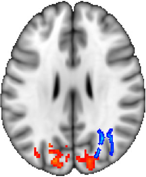
After six weeks of juggling practice, magnetic resonance
imaging detects structural brain changes in the parietal lobe. The image
shows a horizontal cut through the brain to reveal the brain region where
connectivity changed (blue) and the region where neural matter changed
(red).
Our goal was to fill in the missing puzzle pieces and for the first time examine training-related white matter changes by observing the same individuals on multiple occasions and to investigate the relationship between changes in grey and white matter by observing both in the same individuals. Do both neural matter and the brain’s connections change in response to motor training and how are changes in the two brain areas related?
One reason why our findings might have attracted interest beyond the scientific community is that they seem to contradict the common suspicion that learning new skills becomes more difficult with age. In fact, many adults do not attempt to learn an instrument or a new language, arguing that they are "too old" for this. In this respect, revealing the structural flexibility of the adult brain that accompanies the acquisition of a new skill is deeply comforting, because it suggests that learning after childhood might not necessarily be neurobiologically limited.
Further, these findings have sometimes been interpreted as good news for the future treatment of degenerative brain diseases. For example, if measures of brain structure decrease as a result of disease, and training causes an increase, why not combat the ill consequences of the former with the beneficial effects of the latter?
As scientists, however, we need to advise caution. We have yet to understand the nature of experience-related brain changes. Modern imaging methods cannot distinguish changes happening on a cellular level, such as the growth of new neurons and connections, and the growth of new blood vessels and other structures that are extremely important for the efficient functioning of the brain. Further, the relationship between behavioral performance and structural brain changes remains elusive, one notable exemption being Taubert et al. (2010), who studied individuals who had to learn to balance on a tilting board. They found that a measure of balancing performance was related to grey matter of the frontal cortex.
Our work has received attention from the scientific community because it suggests for the first time that in adults, motor training of only six weeks might change the mircrostructure of brain connections underlying the cortex where the neurons of the brain are located. We also found evidence that training might not only change the brain connections, but also the connected cortical brain areas.
This was the first study to use multiple ways to study longitudinal structural changes, i.e. we used diffusion imaging to image white matter and standard structural imaging to image grey matter. Both methods independently found changes that turned out to be in a location near each other. The spatial proximity of changes detected in two independent modalities was exciting at the time. It is now increasingly common to acquire multiple modalities in the same individual. Our work suggests that this approach might reveal inter-related brain processes that manifest in different brain compartments.
![]() Where do you see your research leading in the
future?
Where do you see your research leading in the
future?
The mentioned work and our research have encouraged and will stimulate future research to better understand dynamic structural brain processes. What is the time course of these changes and how do they depend on the task, the training, and the age of the trainees? These are only some of the questions that we will no doubt be examined and hopefully answered in the future. We also found a remarkable variability in the size and direction of the structural changes between individuals. We have yet to understand why these changes differ so much across people, despite the fact that everybody seemed to be learning well.
![]() Do you foresee any social or political
implications for your research?
Do you foresee any social or political
implications for your research?
One question arising from this research for society is the modulation of structural brain changes. If it turns out that measures of brain change are correlated with learning success, then it would only be natural to ask what facilitates these changes. Certain training regimes might be associated with more improvement than others. For example, are training regimes with breaks interspersed better than those without?
Brain imaging might then give us a reason for why some forms of training are better than others, compared to our current understanding which is almost exclusively based on behavioral observations. Finally, these findings might lead to better training regimes that counter-act age-related cognitive decline and facilitate rehabilitation from brain damage.
It is our hope, of course, that ultimately understanding structural brain
changes might benefit healthy as well as ageing individuals and patients.
After all, the functioning of our brain is central to our lives and
well-being.![]()
Jan Scholz
University of Oxford Functional MRI of the Brain (FMRIB) Centre
John Radcliffe Hospital
Headington, Oxford
United Kingdom
KEYWORDS: TRAINING, CHANGES, WHITE MATTER, ARCHITECTURE, VISUAL CORTEX, MYELINATION, SKILLS, RAT.