K. Takahashi, et al.,
"Induction of pluripotent stem
cells from adult human
fibroblasts by defined
factors," Cell,
131(5): 861-72, 30 November
2007. [Kyoto U., Japan; CREST,
Kawaguchi, Japan; Gladstone
Inst. Cardio. Dis., San
Francisco, CA] *243MG
93
1
2
The ENCODE Project Consortium
(
E. Birney, et
al.), "Identification and
analysis of functional elements
in 1% of the human genome by
the ENCODE pilot project,"
Nature, 447(7146):
799-816, 14 June 2007. [80
institutions worldwide] *178FV
68
4
3
Intl. HapMap Consortium
(K.A. Frazer, et al.),
"A second generation human
haplotype map of over 3.1
million SNPs," Nature,
449(7164): 854-61, 18 October
2007. [72 institutions
worldwide] *221LY
61
3
4
A.
Barski, et
al., "High-resolution
profiling of histone
methylations in the human
genome," Cell,
129(4): 823-37, 18 May
2007. [NHLBI, NIH,
Bethesda, MD; U. Calif.,
Los Angeles] *172FA
50
†
5
V. Cherezov, et al.,
"High-resolution crystal
structure of an engineered
human
ß2-adrenergic
G protein-coupled receptor,"
Science, 318(5854):
1258-65, 23 November 2007.
[Scripps Res. Inst., La Jolla,
CA; Stanford U., CA] *233JG
45
2
6
M. Wernig, et al.,
"In vitro
reprogramming of fibroblasts
into a pluripotent ES-cell-like
state," Nature,
448(7151): 318-24, 19 July
2007. [5 U.S. institutions]
*191GC
43
9
7
K. Okita, T. Ichisaka, S.
Yamanaka, "Generation of
germline-competent induced
pluripotent stem cells,"
Nature, 448(7151):
313-7, 19 July 2007. [Kyoto U.,
Japan; Japan Sci. Tech. Agency,
Kawaguchi] *191GC
41
†
8
T.S. Mikkelsen, et
al., "Genome-wide maps of
chromatin state in pluripotent
and lineage-committed cells,"
Nature, 448(7153):
553-60, 2 August 2007. [6 U.S.
institutions] *195XV
39
10
9
S. Vasudevan, Y. Tong, J.A.
Steitz, "Switching from
repression to activation:
MicroRNAs can up-regulate
translation," Science,
318(5858): 1931-4, 21 December
2007. [Howard Hughes Med.
Inst., Yale U. Sch. Med., New
Haven, CT] *243HE
38
†
10
E.S. Lein, et al.,
"Genome-wide atlas of gene
expression in the adult mouse
brain," Nature,
445(7124): 168-76, 11 January
2007. [Allen Inst. Brain Sci.,
Seattle, WA; Baylor Coll. Med.,
Houston, TX; Max Planck Inst.
Biophys. Chem., Goettingen,
Germany] *124QF
One of the delights of scanning the most highly cited papers
is the opportunity to be at least vaguely aware of a huge range of
subjects. One of the drawbacks is that it is impossible to be more
than vaguely aware. Sometimes a paper is so astonishing that it is
difficult not to be struck dumb. Such a paper is at #10.
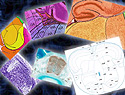
From the Allen Mouse Brain
Atlas. Details
"Genome-wide atlas of gene expression in the adult mouse brain"
beggars belief. It maps, in three dimensions, which of more than
20,000 genes is expressed where in the mouse brain. Not a vague
hand-waving where, like "the neo-cortex," but a precise location
that might be no more than a few cells in volume. A raft of
scientists, mostly at the Allen Institute for Brain Science in
Seattle, adopted an assembly-line approach that integrates several
technologies and where the numbers tell only part of the story. The
effort hinges on an inbred mouse strain that shows minimal variance
across individuals. This is not the absolute cellular determinism
of the nematode worm Caenorhabditis elegans, where each
cell develops in exactly the same way in every non-mutant
individual. The individual mice are, however, sufficiently alike
that the researchers could, as they report, "treat the brain
essentially as a complex
but highly reproducible three-dimensional tissue array."
Not one three-dimensional array but 21,500—one per
gene—plus a reference atlas that holds them all together.
Each mouse brain was sliced into around 130 slices 25 µM
thick in which activated genes were sought using a staining
technique called in-situ hybridization. The automated staining
procedure dealt with the million sections of brain at a rate of
16,000 a week, and each image was then photographed at high and low
magnifications by another automated system to result in 85 million
images. A final system judged the quality of images, which were
then assembled into a three-dimensional viewer that will show the
locations in which a particular gene is expressed and what genes
are expressed in a particular location.
Perhaps the most surprising result is that about 80% of the genes
assayed were expressed in some part of the brain. This is higher
than had been predicted by expression microarray analysis of larger
chunks of brain tissue. It means that many genes are expressed
either at low levels overall or at higher levels but in a small
number of cells. Either way, studies of larger brain regions had
missed them.
The corollary of this is that most genes are
expressed in relatively few cells; 70.5% of the genes are
expressed in fewer than 20% of the cells. The expression
pattern, not surprisingly, reveals something about function.
Among the near-ubiquitous genes, found in all cell types
throughout the brain, are basic housekeeping genes for things
like inter-cellular signalling and general cellular
metabolism. Other genes are associated with particular cell
types. For example, oligodendrocytes, which provide the
insulting myelin sheath around nerve axons, are rich in genes
for lipid synthesis and myelination. And, pleasingly, there is
no evidence for genes one would not expect to be present, such
as those involved in the immune response, meiosis, or blood
coagulation.
Physical regions of the brain also have characteristic patterns of
gene expression. One of the most complex analyses in the
Nature paper looks at the pattern of gene expression in
voxels—the 3-D equivalent of a pixel. The Allen Mouse Brain
Atlas makes it possible to ask which genes are active or suppressed
in each voxel and then to organize the voxels in search of
patterns. If the voxels are grouped according to the large brain
structure they belong to, then there are strong correlations in
expression patterns among voxels in the same structure—no
surprise there—and "complex but distinct correlations"
between brain regions. Group the voxels by their correlations with
other voxels, however, and a different pattern emerges. Large
anatomical regions that are more or less undifferentiated, such as
the cerebral cortex, give a single tight cluster of voxels. By
contrast, structures with discrete anatomically distinct nuclei,
such as the hypothalamus, have smaller local clusters that are
intermingled.
There’s a great deal more neuroanatomical richness in the
paper, which uses gene expression to redefine aspects of brain
structure and which is revitalizing efforts to understand the
brain. Astonishing though its contents are, even more magical is
the publicly available window on the masses of accumulated data. Go
to brain-map.org and I guarantee that unless
you are a neuroscientist (in which case none of this will be
news to you) the richness of the data and the ease with which it
can be manipulated and visualized will combine to occupy hours
of your life. Alas, the impossibility of formulating even a
mildly interesting question to ask of all those data will
reinforce any feelings of inadequacy you might have had on first
reading the paper.
Dr. Jeremy Cherfas is Science Writer at Bioversity
International in Rome, Italy.
KEYWORDS: NEUROGENOMICS, MOUSE BRAIN, GENE EXPRESSION, GENOME-WIDE
ATLAS, ALLEN INSTITUTE FOR BRAIN SCIENCE, IN SITU HYBRIDIZATION,
ALLEN MOUSE BRAIN ATLAS.