Ray M. Kaplan talks with
ScienceWatch.com and answers a few questions about
this month's Emerging Research Front in the field of
Microbiology. The author has also sent along images of
their work.
Article: Drug resistance in nematodes of veterinary
importance: a status report
Authors:
Kaplan,
RM
Addresses: TRENDS PARASITOL, 20 (10): 477-481 OCT
2004
Univ Georgia, Coll Vet Med, Dept Infect Dis, Athens, GA
30602 USA.
Univ Georgia, Coll Vet Med, Dept Infect Dis, Athens, GA
30602 USA.
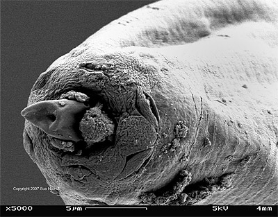
Figure 1 - SEM HC Dorsal lancet: This image
shows a scanning electron micrograph of the
anterior end of a Haemonchus
contortus worm. Clearly visible is the
worm's feeding apparatus, the dorsal
lancet, that enables this blood sucking
worm to feed on blood after piercing the
abomasal (stomach) mucosa. This nematode
has the highest levels of drug resistance
of any parasitic worm, and in some areas it
is resistant to every known anthelmintic
(dewormer) drug.
View
larger image at high
resolution (allow time to
load). Close new browser window to
return to this page.
Figure
2:
Figure 2 - Haemonchus on surface: This
image shows the inside of the abomasum
(stomach) of a goat with a heavy infection
of Haemonchus contortus. Red color
of worms is a result of their feeding on
blood.
View
larger image at high
resolution (allow time to
load). Close new browser window to
return to this page.
Figure
3:
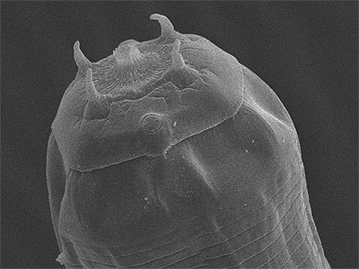
Figure 3 - Clong_ant: This image shows a
scanning electron micrograph of the
anterior end of a Cylicostephanus
longibursatus worm. This species is
the most common nematode parasite of horses
throughout the world, and has been shown to
be resistant to virtual all available
equine anthelmintics.
View
larger image at high
resolution (allow time to
load). Close new browser window to
return to this page.