Xiaoyuan Chen talks with
ScienceWatch.com and answers a few questions about
this month's Fast Moving Front in the field of
Chemistry. The author has also sent along images of
their work.
Article: Peptide-labeled near-infrared quantum dots
for imaging tumor vasculature in living
subjects
Authors: Cai, WB;Shin, DW;Chen, K;Gheysens, O;Cao, QZ;Wang,
SX;Gambhir,
SS;Chen,
XY
Journal: NANO LETT, 6 (4): 669-676 APR 2006
Addresses: Stanford Univ, Sch Med, MIPS, 1201 Welch Rd,
Stanford, CA 94305 USA.
Stanford Univ, Sch Med, MIPS, Stanford, CA 94305 USA.
Stanford Univ, Sch Med, Bio X Program, Dept Radiol,
Stanford, CA 94305 USA.
(addresses have been truncated)
Why do you think your paper is highly
cited?
Coauthor
Weibo Cai
Coauthor
Sanjiv Gambhir
Coauthor
Shan Wang
Even though previous studies have demonstrated the feasibility of using
quantum dots for tumor vasculature targeting, this is the first report of
imaging of tumor vasculature with
quantum dots in a non-invasive manner.
Does it describe a new discovery, methodology, or
synthesis of knowledge?
The conjugation chemistry described in this paper is robust and highly
reproducible, which has become essentially the standard method of
nanoparticle modification. The study addresses several key issues related
to the in vivo application of quantum dots and other
nanomaterials, such as particle size, circulation half-life, and
reticuloendothelial system (RES) uptake.
Would you summarize the significance of your paper
in layman's terms?
This particular peptide nanoparticle conjugate recognizes the new vessels
grown out of a malignant tumor which can be visualized by a specially
designed camera. Such kind of molecular imaging technique will have great
potential in cancer diagnosis, imaging guided surgical tumor removal, as
well as treatment management.
How did you become involved in this research and
were there any particular problems encountered along the way?
Our lab focuses on molecular imaging probe development. We have previously
worked with fluorescent dyes for near-infrared fluorescence optical imaging
studies. However, fluorescent dyes are not bright enough and photostable
enough. Furthermore, fluorescent dyes can produce toxic radicals and
photoproducts upon repeated excitation. It is also rather difficult to
multiplex several colors. Quantum dots, on the other hand, have
size-tunable narrow emission spectra, ideally suited for in vivo
imaging application, but were not previously well studied.
Where do you see your research leading in the
future?
In this particular study we used quantum dots from commercial sources.
These materials contain toxic heavy metals such as cadmium. We are now
trying to develop alternatives to cadmium chalcogenide
nanocrystal emitters. Other than RGD peptides, we
are also testing other targeting molecules including antibodies,
proteins, and peptides.
We are also studying the effect of particle size, rigidity, and surface
chemistry on the targetability and cellular distribution of the newly
developed biocompatible quantum dot conjugates. We are hopeful that our
nanobiotechnology will be clinically relevant and translated into clinical
use in the foreseeable future.
Do you foresee any social or political implications
for your research?
Nanotechnology plays an essential role in molecular
imaging and future molecular medicine. Despite the great promise of
nanomedicine, there are still major hurdles such as biocompatibility,
pharmacokinetics, targeting efficiency, cost-effectiveness, and
acute/chronic toxicity, most of which are virtually untouched. This
paper, along with our other related publications, will likely gain a lot
more interest from the scientific and medical community, helping to push
the field of nanomedicine forward.
Xiaoyuan (Shawn) Chen, Ph.D.
Molecular Imaging Program at Stanford (MIPS)
Department of Radiology, Bio-X & Biophysics
Stanford University School of Medicine
Stanford, CA,
USA Web
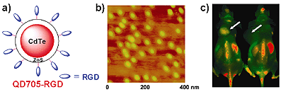
RGD peptide-conjugated QD705 for NIRF imaging of tumor vasculature. a) A
schematic illustration of the probe QD705-RGD. b) An atomic force
microscopy image of QD705-RGD deposited on a silicon wafer. c) In vivo NIRF
imaging of tumor vasculature in U87MG human glioblastoma tumor-bearing
mice. The mouse on the left was injected with QD705-RGD and the mouse on
the right was injected with QD705. Arrows indicate tumors.